Picture Of Forearm Muscles And Tendons / The Anatomy Of The Elbow : The extensor digitorum is a muscle belly, passing first into four tendons, which in turn transformirovalsya in stretching the tendon fixed to the base of the distal.
byErik Morris•
0
Picture Of Forearm Muscles And Tendons / The Anatomy Of The Elbow : The extensor digitorum is a muscle belly, passing first into four tendons, which in turn transformirovalsya in stretching the tendon fixed to the base of the distal.. Anterior, lateral or posterior compartment. A square shaped muscle found deep to the tendons of the fdp and fpl. The 3 muscle groups of the forearm each have their own unique form. The posterior compartment of the forearm (or extensor compartment) contains twelve muscles which are chiefly responsible for extension of the wrist and digits, and supination of the forearm. 12 (4 superficial + 3 mobile wad + 5 deep).
Most of these originate from the lateral epicondyle. All superficial muscles are arises from the medial epicondyle of humerus but they are inserted into the different part except. The superficial group arises mostly from the posterior aspect of the lateral epicondyle of the humerus by a common tendon. Back when sniffing tobacco was cool. A deep layer , intermediate layer and superficial layer.
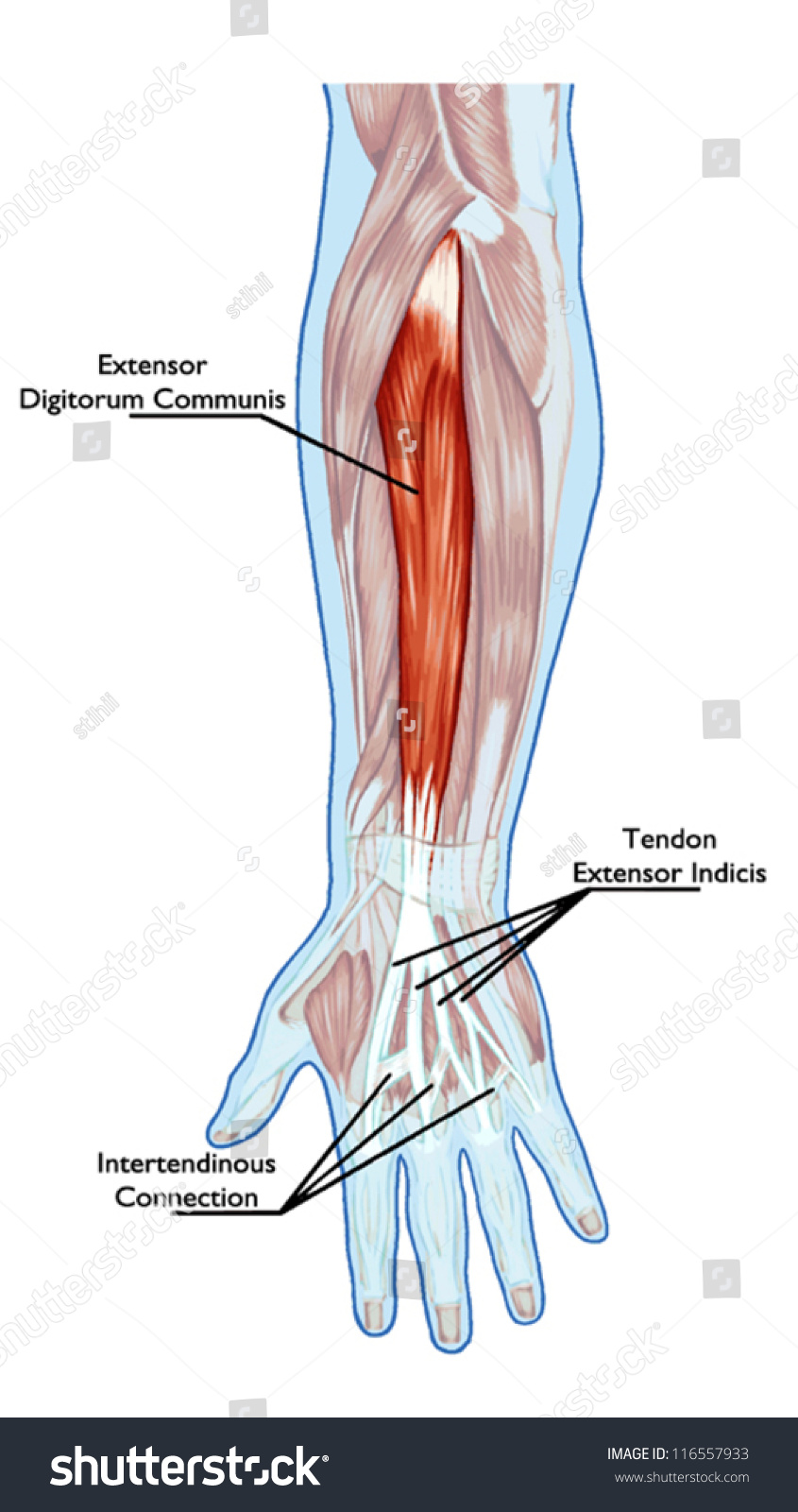
Anatomy Muscular System Hand Forearm Palm Stock Vector Royalty Free 116557933 from image.shutterstock.com Most of these originate from the lateral epicondyle. This type of forearm grade iii strain of forearm muscle: In the anterior compartment, they are split into three categories: Antagonist of forearm flexors ( bra… flexion powerful of elbow and supination of forearm; It turns… inflamed common flexor tendon cft. The muscle fibers then descend towards the wrist area where they converge onto a narrow tendon. 12 (4 superficial + 3 mobile wad + 5 deep). The forearm muscle strains are graded into three categories which are described below:
This retinaculum prevents bow stringing of the tendons when the flexor muscles contract and also help improve the effective of the muscles by changing the.
Find stockbilleder af forearm muscles tendons i hd og millionvis af andre royaltyfri stockbilleder, illustrationer og vektorer i shutterstocks samling. A deep layer , intermediate layer and superficial layer. The 3 muscle groups of the forearm each have their own unique form. The muscle fibers then descend towards the wrist area where they converge onto a narrow tendon. Do it yourself as shown in the picture! Tusindvis af nye billeder af høj kvalitet tilføjes hver dag. The forearm muscle strains are graded into three categories which are described below: 12 (4 superficial + 3 mobile wad + 5 deep). Also, pollicis means thumb in latin. These types of strains are quite severe and involve complete rupture of the muscle fibers and tendons. Grade i strain of forearm muscle: The superficial group arises mostly from the posterior aspect of the lateral epicondyle of the humerus by a common tendon. The forearm is the region of the upper limb between the elbow and the wrist.
In the anterior compartment, they are split into three categories: They receive additional fibers from the deep fascia of the forearm near the elbow, and from the septa which pass from this fascia between the individual muscles. A common muscle belly is shared by all the fingers. This type of forearm grade iii strain of forearm muscle: Grade i strain of forearm muscle:
Posterior Forearm Muscles Ankiweb from dl8.ankiweb.net Do it yourself as shown in the picture! Find stockbilleder af forearm muscles tendons i hd og millionvis af andre royaltyfri stockbilleder, illustrationer og vektorer i shutterstocks samling. The pronator teres has two heads of. They receive additional fibers from the deep fascia of the forearm near the elbow, and from the septa which pass from this fascia between the individual muscles. The tendons travel down the forearm through a tough band of tissue on top of the wrist. You've got these four tendons coming off. In the anterior compartment, they are split into three categories: Edc tendons straighten the index, middle, ring and small fingers.
Two of the tendons combine visually so the end result looks like two thick tendons, with a long hollow between them.
Most commonly it is the tendon of the extensor carpi radialis brevis muscle that is weakened or torn from injury or overuse. Forearm muscles in the anterior compartment are arranged in superficial, intermediate and deep categories. Two of the tendons combine visually so the end result looks like two thick tendons, with a long hollow between them. Edc tendons straighten the index, middle, ring and small fingers. It is separated from the anterior compartment by the interosseous membrane between the radius and ulna. The muscle fibers then descend towards the wrist area where they converge onto a narrow tendon. The muscles of this group take origin from the medial epicondyle of the humerus by a common tendon; The tendons travel down the forearm through a tough band of tissue on top of the wrist. Flexor digitorum profundus tendons (long flexor tendons share the same synovial sheath, referred. In the anterior compartment, they are split into three categories: Learning their anatomy will help you design awesomely dynamic arms. 12 (4 superficial + 3 mobile wad + 5 deep). There are many muscles in the forearm.
You've got these four tendons coming off. Muscles of the forearm segregate into these compartments consisting of (1) an anterior group (the flexors seven superficial and five deep muscles occupy the posterior forearm. Edc tendons straighten the index, middle, ring and small fingers. And if we follow it down the forearm, you can see it gives off four tendons. Lesson on the anatomy of the forearm:
1 from All superficial muscles are arises from the medial epicondyle of humerus but they are inserted into the different part except. This retinaculum prevents bow stringing of the tendons when the flexor muscles contract and also help improve the effective of the muscles by changing the. Two of the tendons combine visually so the end result looks like two thick tendons, with a long hollow between them. The 3 muscle groups of the forearm each have their own unique form. You've got these four tendons coming off. The posterior compartment of the forearm (or extensor compartment) contains twelve muscles which are chiefly responsible for extension of the wrist and digits, and supination of the forearm. This type of forearm grade iii strain of forearm muscle: And if we follow it down the forearm, you can see it gives off four tendons.
The posterior compartment of the forearm (or extensor compartment) contains twelve muscles which are chiefly responsible for extension of the wrist and digits, and supination of the forearm.
Cross sectional anatomy of the upper limb : Learning their anatomy will help you design awesomely dynamic arms. The muscle fibers then descend towards the wrist area where they converge onto a narrow tendon. Lesson on the anatomy of the forearm: A square shaped muscle found deep to the tendons of the fdp and fpl. Tusindvis af nye billeder af høj kvalitet tilføjes hver dag. Originates from the anterior surface of the ulna and attaches to the. See anatomy pictures of the 27 bones in the hand and wrist, how they are connected with tendons and muscles and the nerves that run through the skeletal structure. The muscles of this group take origin from the medial epicondyle of the humerus by a common tendon; This type of forearm grade iii strain of forearm muscle: It turns… inflamed common flexor tendon cft. While the ventral side of the forearm is not exactly less complicated than the dorsal side, it appears less complicated on the. Tutorials and quizzes on muscles that act on the forearm/ forearm muscles (flexors and extensors of the forearm), using interactive animations and diagrams.
When identifying the function of the forearm muscles, it is important to note that any forearm compartment muscle that crosses the elbow joint will act at this joint picture of forearm tendons. The forearm is the region of the upper limb between the elbow and the wrist.